


|
What is Retinitis Pigmentosa?
Have you experienced night vision loss? Have you experienced loss of your Peripheral Vision? Has your eye doctor said that you have scars in the back of your eye (Retina)? You may have Retinitis Pigmentosa. RP affects photoreceptors (specialized cells that are sensitive to light) and the Retinal Pigment Epithelium (RPE). The RPE is located between blood vessels of the choriocapillaris and the light-sensitive outer segments of the photoreceptors. The RPE closely interacts with photoreceptors in the maintenance of visual function. 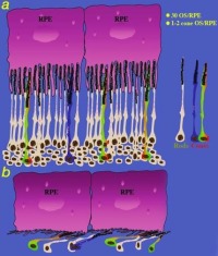
Most of the mutations seen in Retinitis Pigmentosa lie in genes expressed in rod photoreceptors (which are responsible for night vision). This is why RP typically starts with Island Scotoma (blind spot) in the Peripheral Vision and progresses to ring-shaped Scotoma. In rare cases the gene mutation may be expressed in cone photoreceptors (which are responsible for sharp central vision and Color Vision). In the very small central visual field left in advanced cases, some retain good function throughout the lifetime while others begin to degenerate, resulting in total blindness. What are the signs and symptoms of Retinitis Pigmentosa?
Retinitis Pigmentosa as noted above describes a variety of hereditary dystrophies of the Retina which have in common the following signs and symptoms: Characteristic Retina Findings such as: 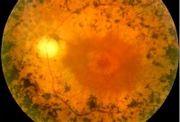
How is Retinitis Pigmentosa diagnosed?
Because the majority of diseases classified as RP usually have a significant period of no symptoms following birth. The onset of symptoms can range from early childhood to late adulthood, depending on the genetic type. During this period of no symptoms, diagnostic testing of affected individuals can potentially be achieved by a routine eye examination, electroretinography (ERG) or DNA testing. ERG is an objective measurement of retinal function that can be used to monitor for deterioration. A routine eye examination may reveal early Retinal Pigmentary changes or subtle abnormalities of the Macula. There are several ways of monitoring the function of the Macula including: There are several ways of monitoring the function of the Macula including: 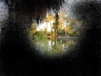
How is Retinitis Pigmentosa managed or treated?
If you are asymptomatic (that is, you have no complaints), results of the diagnostic testing may be difficult to interpret. Early changes may be so mild that counseling you of your potentially blinding disease must be handled with caution. Even objective ERG testing may be difficult to interpret, particularly if you have high Myopia. Children who are not cooperative with the testing also make it hard to interpret findings. If you are given that diagnosis, you may be overwhelmed by the news. Just like is the case with other vision debilitating diseases, you may be encouraged to speak to a mental health provider to help you adapt to the prospect. One of your first priorities is to get the best spectacle prescription to maximize your vision. Also, maximize your functional vision by consulting a Low Vision specialist. Many devices are available to help patients with night vision difficulties. The next priority in managing RP is to have annual eye examinations, including visual field testing and periodic (such as every 2 to 3 years) ERG evaluations. Changes in examination findings over the course of time can uncover progression of the disease and can help in determining what options to deal with the remaining functional vision. In addition to monitoring for progression of the disease, annual eye examinations will allow your eye doctor to determine if you have Cataracts (a clouding of the crystalline lens of your eye) and refer you for Cataract surgery when necessary. There is no effective treatment for RP. Some studies have suggested that treatment with antioxidants may slow the disease. However, this treatment must be weighed against the uncertain risk of long-term adverse effects from large chronic doses of Vitamin A. Annually check your liver enzymes if you choose this option. Another antioxidant that has been studied for the slowing down of the progression of Retinitis Pigmentosa is Docosahexaenoic acid (DHA). DHA is an omega-3 polyunsaturated fatty acid and antioxidant. For complications such as Macula Edema, a drug called Acetazolamide has shown the most encouraging results with some improvement in visual function. Adverse effects, including fatigue, kidney stones, loss of appetite, hand tingling, and anemia may limit its use. Microchip implants that go inside the retina and act like a microscopic video camera are in the early stages of development for treating blindness associated with serious eye conditions such as Retinitis Pigmentosa. |