


|
Peripheral Vision
Have you noticed that you hit things off to your side because you cannot see them? You may feel that your central vision is good, but do you miss things either to your left or right when you are walking? You may have experienced a loss in your Peripheral Vision. What does loss of Peripheral Vision mean?
Loss of Peripheral eyesight may occur as the result of a variety of eye diseases or trauma. The normal Visual Field (VF) extends more than 90 degrees temporally (on the side of the ears), 60 degrees nasally (on the side of the nose) and superiorly, and about 70 degrees inferiorly. 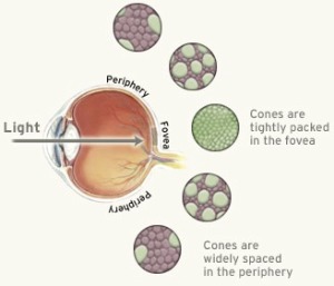
When you look at the diagram, you can see a graphical depiction of the VF of a normal right eye and how it translates to vision compared to one of a right eye with Tunnel Vision. 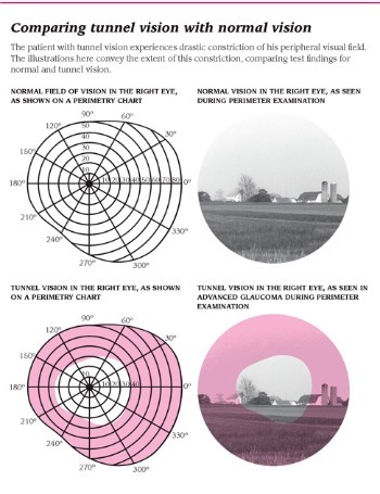
The surface of the Retina receives an image of objects in the VF much like photographic film records a landscape. The nerve fibers from the Retina are arranged in a pattern that is repeated in the visual pathway from the Optic Nerve through to the cortical visual center in the occipital lobes of the brain. Any defect or abnormality of the VF may, through correct interpretation, reflect disease or damage to a specific portion of the visual pathway. Not all loss of Peripheral Vision, however, is due only to Retina or visual pathway disease.
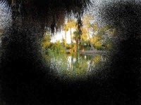
Observe that the child’s right upper lid in the picture below is partially obstructing his Superior Peripheral Vision. If you have loss of your Superior Peripheral Vision, you may see the world as depicted in the image below. 
If your eye doctor documents that your drooping lid is significantly obstructing your vision, then you may be able to have Ptosis surgery covered by your health insurance company since it would not be done solely for cosmetic reasons. The following eye and medical conditions are some of possible causes of Tunnel Vision: Other types of loss of Peripheral Vision can be seen in: How is loss diagnosed?
The diagnosis of loss of Peripheral or of Tunnel Vision is primarily going to depend on your complaints or symptoms. For example, Depending on the issues that you bring up while talking to your eye care provider, he or she will decide whether you need further testing of your VF. Most routine eye exams will include some type of VF screening test. The most common and widely used is Confrontation Testing because it is a rapid screening VF test. Your eye doctor will have you count fingers in the periphery to uncover any areas you have trouble seeing. It is suitable for identifying large defects in the Peripheral Vision and less ideal for finer testing. Many eye doctors have computerized screening VF tests which are more sensitive than Confrontation Testing. However, for more quantitative data, computerized threshold VF testing is best. This type of VF testing will measure in units of decibels the sensitivity throughout your Tunnel Vision and compare it to people of your age to see if your sensitivity is average or below average (indicating disease). These numbers can then be compared to future testing results to determine if your condition is getting worse or staying stable. How is loss managed or treated?
Once the reason why you have experienced loss of Peripheral Vision is discovered, the treatment will be tailored to find and treat the underlying cause. Once the cause is treated, your eye care provider can refer you to a Low Vision specialist to help you function better with your remaining Vision. Because Tunnel Vision will make it more difficult for you to navigate through the environment, your Low Vision specialist will prescribe optical devices and provide training that will improve your awareness of the environment and independent travel ability. Once your eye doctor explains to you the extent of your Tunnel Vision, your ability to compensate for the loss will be explored. Mobility evaluation by a certified orientation and mobility specialist may also be recommended. Your Low Vision specialist may provide you with one or more of the options below: Use of any of these optical devices will require training before any true potential for success can be determined. You may also want to consult a mental health provider if you find that your Tunnel Vision is progressive and making you feel depressed. |
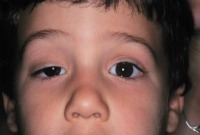 Suppose you have loss of your Superior Peripheral Vision. This may be due to the physical obstruction of a drooping upper lid known as Ptosis.