


|
Color Blind Test
Has your family told you on multiple occasions that your clothes do not match in color? Or have you noticed that your child’s clothes consistently do not match in color? Are you unable to tell a difference in the color of traffic lights? You (or your child) may need to go to an eye doctor and have a Color Blind Test. What should you expect from a Color Blind Test?
A Color Blind Test will help your eye doctor determine if you are able to distinguish between colors. If you are only able to discriminate certain colors and not others, then you have a Color Deficiency. If you are unable to appreciate any color at all, then you have Color Blindness. Because color vision ability can vary in severity and can be deficient in certain colors and not others, a Color Blind Test will be used to determine the type, and in some cases, the severity of Color Deficiency. A Color Blind Test may be qualitative (i.e., can you see color and if not, which ones can you not see?) or quantitative (i.e., how severe is your Color Deficiency?). Thus, the quantitative tests try to provide a measure of the severity of Color Deficiency. Regardless of which Color Blind Test is used, there are factors that affect test performance such as: What should you expect from a qualitative Color Blind Test?
The most commonly used screening tests for Color Deficiency in clinical practice are Plate Tests or Pseudoisochromatic Plates (PIP). These tests can be used on children and illiterate adults. Most of these plate tests provide very efficient screening of Congenital red-green defects. Plate tests generally consist of a series of cards on which is printed a number or pattern in multiple colors against a multi-colored background. The figure is easily seen by color normal individuals, but if you have a Color Deficiency, you may have trouble seeing it. Do you see the number 2 in the circle?
A second variety of the Plate test is PIP II, which is used for identifying Acquired Color Deficiency since it tests for blue-yellow defects, more readily seen in acquired eye diseases or medication toxicity. Despite their popularity and advantages, Plate tests have distinct limitations such as: What should you expect from a quantitative Color Blind Test?
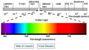
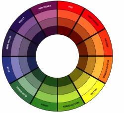
A Color Blind Test category that includes a scoring system is that of Arrangement Tests. These tests were designed by the Navy Commander Dean Farnsworth for military use in 1943. These tests were designed to evaluate color discrimination ability and to classify those tested into groups of Protan, Deutan, and Tritan based on the error axis of the scored results. The basic design of these tests is to present you with colored caps of fixed saturation (how different cap color is from white) and value (refers to relative lightness or darkness of color) selected from the Hue Circle. Newton joined the “orange red” and “violet” ends of the spectrum to create a Hue Circle, and thus shows the spectrum as a continuous gradation of hues from orange red to blue violet, but now the ends are joined by mixed colors red, violet red, red violet, and violet.
You will be asked to arrange randomly placed caps in what you perceive to be a natural order. The color differences between adjacent caps on the Farnsworth-Munsell 100-Hue Test (FM 100-Hue Test) were designed to be very small. The Farnsworth Panel D-15 was designed to have larger color differences. 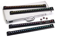 Your eye doctor will score the test by comparing your arrangement of the caps to the correct arrangement, which is indicated by the numbers written on the back of each cap.
 A total error score can be computed for the test and it indicates your aptitude for hue discrimination. A higher score suggests worse discrimination. Your arrangement of caps can also be plotted in order to identify zones or poles of discrimination loss. 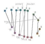
Similarly to the Plate tests, the Arrangement tests have their drawbacks: Another Color Blind Test category that includes a scoring system involves the use of an instrument called an Anomaloscope. This instrument provides a superior clinical method of diagnosing and classifying Color Deficiency. You will be asked to view through the eyepiece to see a circle of color, one half of which is yellow light and the other half is a mixture of red and green light. You will be asked to match the color and brightness of the two halves using two knobs. The amount of each color that you use to match the two halves can indicate the particular kind and severity of Color Deficiency you have. 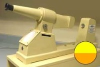
While this instrument is the benchmark color matching test for diagnosis for Red-Green Deficiency, it has several disadvantages as well: A promising new Color Blind Test is a contribution of military research studies and is called the Cone Contrast Test (CCT). The CCT is inexpensive enough to be used in mainstream clinics, but sensitive and specific enough to diagnose Color Deficiency as well as determine the type and score the severity of it. The CCT can be useful in diagnosing Congenital Color Deficiency as well as in monitoring the progression of Acquired Color Deficiency, which can make an impact in your eye doctor’s ability to manage such diseases as Diabetic Retinopathy and Glaucoma. Each Cone type is tested separately in decreasing levels of contrast in order to quantify any loss and the type of loss. This test will be available in automated form and the computer will generate a score for each Cone type. With a measure each visit, you can see how any decrease in color vision sensitivity will be easier to detect. |
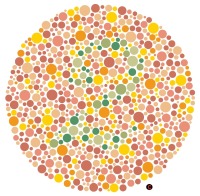 As you can see from the image, the figure and background are drawn in dots. The size of the dots used in plate design can be varied or uniform, but the only difference between the figure and the background is color.